Sunday, May 3, 2015

Determination of Antibiotic Resistance or Sensitivity in Our Unknown Soil Microbe

This week's lab focused on our unknown soil microbe's reaction to antibiotic exposure.

As you probably already know, antibiotics are extremely important in modern medicine. Throughout history, millions of people have died from bacterial infection, and many millions more have been saved because of the discovery of antibiotics. Antibiotics are not effective against viral infections and can work by killing microbes or preventing/inhibiting their growth. Some, such a penicillins and cephalosporins, operate by damaging/compromising the cell wall and/or membrane of a particular bacterial cell. They are also able to inhibit particular enzymes that are necessary for the function of a bacterial cell. Other types affect a bacterial cell's ability to synthesize proteins and are referred to as bacteriostatic.

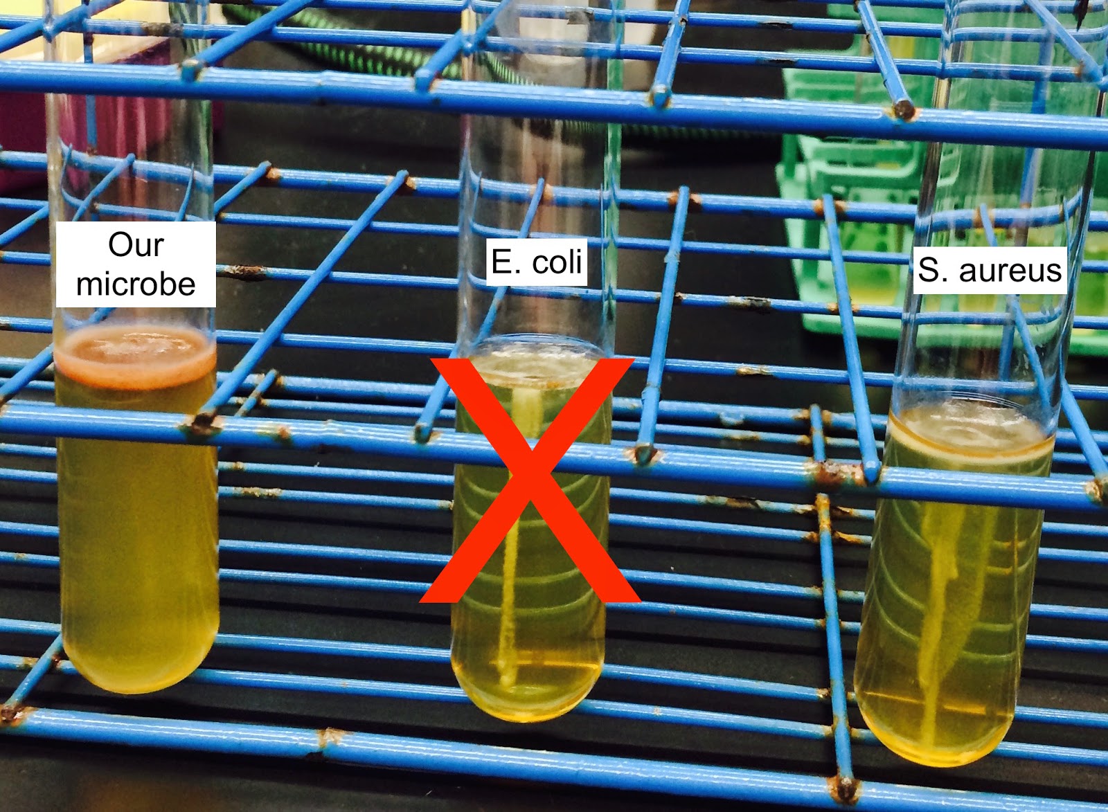
In this week's lab, several bacterial controls and our unknown microbe were plated so that a they grew "lawns." Before incubation, several antibiotic disks of varying types and concentrations were placed on each plate (see photos below). Later, they were each observed to determine whether they were sensitive (unable to grow in the presence of the antibiotic) or resistant (able to grow in the presence of the antibiotic).


K. pneumonia response to Tetracycline, Ampicillin, Carbenicillin, Arithromycin
S. aureus response to Tetracycline, Ampicillin, Carbenicillin, Arithromycin
E. coli response to Tetracycline, Ampicillin, Carbenicillin, Arithromycin
Unknown soil microbe response to Tetracycline, Ampicillin, Carbenicillin, Arithromycin

Antibiotic Sensitivity

Microbes Tested
Tetracycline [30]
Ampicillin [10]
Carbenicillin [100]
Azithromycin [15]
K. pneumonia
Sensitive
Resistant
Sensitive
Sensitive
S. aureus
Sensitive
Sensitive
Sensitive
Sensitive
E. coli
Sensitive
Sensitive
Sensitive
Sensitive
Unknown
Sensitive
Resistant
Resistant
Sensitive

So what is the classification of our unknown microbes? Well using the results from previous weeks, we still believe that our unknown microbe is either clostridium, mycobacterium, or nocardia.

With further research into the possible identifications of our microbe, we discovered that the three possible classifications are categorized as producing beta lactamase. These enzymes break down the antibiotics making the microbes resistant, especially to penicillin-like antibiotics.  This information is also supported by the antibiotics that our microbe is sensitive to because these antibiotics are not beta-lactamases.

To further verify our microbe, we would consider reconducting the acid-fast stain to confirm that we did the procedure properly and support our initial prediction of our microbe being mycobacterium or nocardia. An additional test to determine if our sample is clostridium would be to perform a starch hydrolysis test. This would allow us to determine if our sample is amylase-positive or negative. If amylase-positive, this would support our microbe's identification as clostridium.

After learning so much about our soil microbe, we recommend that you beware of the unique but dirty little creatures hiding all over our campus.

Thanks for tuning in all semester long!
Austin and Anne

http://en.wikipedia.org/wiki/Antibiotics
Mechanisms of beta-lactam resistance in anaerobic bacteria.
Beta-Lactamase Production and Resistance to Beta-Lactam Antibiotics in Nocardia
Genetic analysis of the beta-lactamases of Mycobacterium tuberculosis and Mycobacterium smegmatis and susceptibility to beta-lactam antibiotics.
http://www.austincc.edu/microbugz/starch_hydrolysis.php
http://www.uwyo.edu/molb2210_lab/info/biochemical_tests.htm#spirit_blue


Monday, April 20, 2015

It Doesn't Happen Only in Transylvania: Bacterial Hemolysis and Our Unknown Microbe

In order to further characterize our unknown microbe, this week's lab focused on determining whether or not it is able to lyse red blood cells. 

There are two ways that bacteria are able to lyse red blood cells (RBCs). Some bacteria contain a special compound composed of lipids and proteins known as hemolysin that allows them to form pores in the phospholipid bilayer of the cell membrane of a red blood cell. Other bacteria possess a different type of hemolysin that allows them to enzymatically cleave the red blood cell's phospholipid membrane. 

Bacteria fall into one of three categories: alpha, beta, or gamma. These designations describe their ability to lyse RBCs. Alpha bacteria do not lyse RBCs fully. Because the RBC cell membrane remains intact, they are said to partially break down the cells, and reduce hemoglobin to another chemical that is found in blood agar medium (5% sheep blood). These typically appear as a bruise-like color when plated. Beta bacteria are able to completely lyse RBCs, resulting in a clear "halo" around a plated colony. Gamma bacteria do not interact with red blood cells, and form colonies with no surrounding discoloration on blood agar plates.

Because virulence refers to the pathogenicity of a microbe, we believe that hemolytic bacteria would be more virulent than non-hemolytic bacteria since they would more easily affect/harm an organism that possesses RBCs. Additionally, we would not expect a "typical" soil microbe to be capable of hemolysis because plants and other soil organisms do not contain red blood cells or hemoglobin. Therefore, it would be an unnecessary attribute for them to possess. 

As seen in the photos below, we compared our unknown microbe to other bacteria. We believe our microbe would be categorized as alpha because of the greenish brown discoloration observed on the plate. 


We are still in the process of trying to narrow down the possible identities of our unknown soil microbe.  From all of our lab analyses, this is what we have determined about our microbe:
  • gram-positive
  • acid-fast
  • ferments lactose and/or sucrose
  • catalase positive
  • endospore forming
  • reduces nitrate to nitrite
  • motile
  • alpha hemolysis

Using the dichotomous key provided by Dr. Hanson, we have concluded that our microbe could be any of the following: Mycobacterium, Nocardia, Clostridium.

Check back next week for more details on our microbe.
-Anne and Austin

http://www.microbelibrary.org/component/resource/laboratory-test/2885-blood-agar-plates-and-hemolysis-protocols
http://en.wikipedia.org/wiki/Hemolysin
http://www.encyclopedia.com/doc/1G2-3409800092.html
https://www.wisc-online.com/learn/career-clusters/health-science/mby4307/blood-agar-and-hemolysis

Sunday, April 12, 2015

Need a Fix? Nitrogen Fixation and Reduction in Our Unknown Soil Microbe

      In order to further characterize our unknown soil microbe, this week's lab focused on the process of nitrate reduction. This process is an integral part of the larger nitrogen cycle that alters atmospheric nitrogen into a usable form by plants and back into its atmospheric form. This process can be seen as a balancing act between the nitrogen that is "fixed" by other species of bacteria the nitrate that is removed from the soil by "denitrifying" bacteria and accounts for the majority of the nitrogen that enters the atmosphere. They are essential for the prevention of excessive levels of soil nitrate, which is important for public health and prevent eutrification (an overabundance of nutrients) of water sources.

      Only certain species of bacterial microbes have evolved the ability to reduce nitrate. This process typically occurs in locations that do not provide enough oxygen for aerobic respiration. Though nitrate reduction provides a microbe with a source of energy, it does not produce nearly as much energy as oxygen does in aerobic respiration. Thus, it would make sense that only bacterial species that are commonly found in oxygen-defficient locations would evolve the ability to reduce nitrate.

     We were able to use a simple test to determine whether our unknown microbe was capable of reducing nitrate. Three samples were inoculated with E. coli, P. aergoginosa, and our unknown sample, respectively, along with a control that did not contain a microbe. Samples were incubated for 48 hours. A smaller tube, known as a Durham tube, was located within the inoculated sample tube. If a bubble formed in the top of the Durham tube, it would indicate that our microbe was able to reduce nitrate (NO3) to its gaseous atmospheric state (N2). As seen in the photo below, this was not the case for our microbe. If the microbe were able to reduce nitrate to another form, such as nitrite (NO2), we would see the solution turn red after adding a solution of sulfanilic acid and alpha-naphthylamine to the inoculated sample. After adding the solution, our inoculated sample turned red (bottom photo). Thus, we concluded that our microbe was able to reduce nitrate to nitrite.
Results after 48 hours. Bubble formed in Durham tube in P. aeroginosa only.

Result following addition of sulfanilic acid and alpha-naphthylamine.


Up to this point, the dichotomous key has indicated that our unknown bacterium is Clostridium. According to the internet sources listed below, this identification still holds true with the results of this week's experiment. Come back next week for more details!!

http://en.wikipedia.org/wiki/Nitrogen_fixation
http://en.wikipedia.org/wiki/Denitrifying_bacteria

- Austin

Monday, April 6, 2015

Check Out Our Microbe's Moves: An Analysis of Our Soil Microbe's Motility

This past week in lab, Austin and I discovered another feature about our little microbe. We used the soft agar deep test to determine if our microbe is motile or not. For the experiment, we used a motile control (E. coli) and an immotile control (S. aureus). However, the E. coli sample did not behave properly because the results indicated that the sample was immotile, which is not true. As seen in the picture below, our microbe was very motile and moved all throughout the agar, which was determined by the cloudiness of the sample. A thick layer of microbes also developed on top of the agar.
Soft agar deep test results
So what makes bacteria motile or immotile? The main structural feature that allows microbes to be motile is the presence of flagella, which can vary in number and arrangement. As we studied in cell and molecular biology, flagella are anchored to cells by a hook that attaches to the basal body. A rotary motor unit does the actual whipping of the tale and can vary in speeds from 200 to 1000 revolutions per second, which is crazy fast!
There are advantages for microbes to evolve to be motile. The presence of flagella allows bacteria to swim towards nutrients and dodge harmful substances. They can also swim to new areas and colonize. Many bacteria are motile, however, some bacteria are immotile and have evolved to stay that way. A reason for remaining immotile is dependent on the types of fluid the bacteria travel through. Flagella are not useful when moving through viscous media and microbes that live in these environments do not want to be motile.

Each week in lab, we are continuously getting closer to identifying our microbe. To ensure the accuracy of our endospore verification test from last week's lab, we reran the experiment. The results ended up being the same but this time the controls produced the expected results. Below is a new picture of our endospore results.
Results from endospore verification redo
Hopefully next week, we will be able to narrow down the possible identifications of our microbe.
Check back next week for an update from Mills!
-Anne


Sunday, March 29, 2015

The Quick and Dirty: Endospore Verification in Our Unknown Bacterium

       Several types of bacteria are known to form tiny versions of themselves when the going gets tough. These structures are referred to as endospores, and allow a bacteria to preserve itself when it encounters an environment that is unfavorable for reproduction or is lacking in nutrients. These minuscule spores contain a replicated version of the cell's genetic material, several types of ribosomes, and a chemical known as dipicolinic acid, which helps the endospore remain in its dormant phase. Endospores are extremely difficult to destroy or "kill." They can survive the most extreme of conditions: extremely high or low temperatures, ultraviolet radiation, extremes in pH, and even excessive drying. What's more, endospores of some species can survive for centuries or even millennia (hopefully nowhere near my house)! The evolutionary acquisition of bacterial endospores was probably favored in species that were often exposed to nutrient-lacking or high stress environments. Some bacterial cells may not have not evolved the ability to produce endospores simply because they did not often encounter these types of environments and, thus, it would not have been energetically favorable to do so.

     In this week's experiment, we performed an endospore stain to further narrow the possible identities of our microbe. Cells lacking endospores should appear light pink in color following the stain. Cell containing endospores should contain darker blue structures much smaller than the larger pink cells when examined under a microscope. Because endospores are able to tolerate extremes in temperature, we also performed a heat shock on a sample of our unknown bacterium. If growth was observed several days following the heat shock, it would indicate that endospores were present in the sample.

     As seen in the picture to the right, growth was observed in both the heat shocked sample (right tube) and the control sample (left tube) and four days of incubation, indicating the presence of endospores in our unknown bacterium. When viewed under a microscope, we saw tiny blue/purple structures scattered across the slide among the larger bacterial cells. These were assumed to be endospores. According to the dichotomous key provided, this opens the possibility that our bacterium could be Clostridium.

http://www.biomedcentral.com/content/pdf/1471-2164-13-265.pdf
http://en.wikipedia.org/wiki/Endospore#Formation_and_destruction

Come back next week for more dirty details!
- Austin

Monday, March 23, 2015

Catalase, Slants, and Butts: A look into our soil microbe's catalase activity and carbohydrate metabolism


In lab, the week before Spring Break, Austin and I further analyzed our soil microbe using the catalase activity test and triple sugar iron test. The overall premise of the catalase activity test is to determine if the microbe is catalase positive or negative. Catalase is an antioxidant enzyme that protects cells from oxidative stress. In this case, if catalase is present, then it will neutralize the hydrogen peroxide into water and oxygen gas. This neutralization of catalase-positive samples causes bubbling after adding a drop of hydrogen peroxide, and catalase-negative remains unchanged. Some microbes have evolved to acquire catalase activity. This could be primarily due to the added protection from oxidative stress that catalase-positive microbes possess.

To determine the catalase activity of our soil microbe, we first observed the reactions of a catalase-positive and catalase-negative control. When we added hydrogen peroxide to our soil microbe, the sample bubbled indicating it was catalase-positive.

The other test we conducted was the triple sugar iron test, which is used to determine the type of carbohydrate metabolism that the microbe utilizes. This specific experiment differentiates between lactose, glucose, and sucrose fermentation and hydrogen sulfide production. We inoculated our soil microbe and 4 controls on slants containing the triple sugar iron agar. After returning to the lab after 2 days of incubation, the controls behaved as we anticipated. The controls were characterized based on the color of the slant and butt. Our soil microbe was yellow at the slant and butt. This color is characteristic of an acid over acid tube, which we also saw with the E. coli control. Metabolically, our soil microbe fermented lactose and/or sucrose. Our soil microbe and the controls can be seen in the picture below.

(FROM LEFT TO RIGHT) Our soil microbe, B. megaterium, E. coli, P. areugrinosa, P. vulgaris
The fact that our soil microbe is catalase-positive and a lactose and/or sucrose fermenter does not change any of the previous classifications of our microbe.  It is still narrowed down to either genus Myobacterium or Nocardia.


Check back next week for more information from Austin about our soil microbe's endospore stain!